

Immune Checkpoint Inhibitors
The immune checkpoint inhibitors are antibodies against cytotoxic T-lymphocyte-associated antigen 4 (CTLA-4) and programmed cell death protein-1 (PD-1), which regulate immune responses at different points in the immune response.
CTLA-4
- CD28 is a co-stimulatory receptor expressed on T cells, and CTLA-4 is a coinhibitory receptor expressed on T cells.
- CD28 and CTLA-4 share identical ligands: B7-1 (CD80) and B7-2 (CD86), but CTLA-4 has a much higher affinity for these ligands than CD28.
- Naïve and memory T cells express CD28 but not CTLA-4.
- When antigen-MHC complex binds to the T cell receptor, CTLA-4 is upregulated in the secondary lymphoid organs where T cell activation occurs.
- The level of CTLA-4 expression depends on the amplitude of the initial TCR signaling. The stronger the stimulation through the TCR and CD28, the greater the amount of CTLA-4 that is expressed.
- CTLA-4 has three main functions:
- It dampens the activation of T cells by binding to B7-1 and B7-2 and preventing CD28 from being able to bind these ligands.
- It delivers inhibitory signals to T cells which downmodulate the amplitude of T cell responses.
- It is expressed on T regulatory cells constitutively and also increases the immunosuppressive activity of Tregs. Therefore, and antibodies blocking CTLA-4 will allow B7 ligands to bind CD28, ehnahce the amplitude of T cell responses, and prevent Tregs from exerting their immunosuppressive effects. Together, these effects promote anti-tumor immunity.
PD-L1
- Unlike CTLA-4, which acts in the secondary lymphoid organs at the time of T cell activation, PD-1 acts in the tumor microenvironment where T cells encounter an infection or tumor.
- Activated T cells upregulate PD-1 and continue to express it in the peripheral tissues. Cytokines such as IFN-gamma induce the expression of PD-L1 on epithelial cells and tumor cells. PD-L2 is expressed on macrophages and dendritic cells.
- The main role of PD-1 is to limit the activity of effector T cells in the periphery and prevent excessive damage to the tissues during an immune response.
- Chronic antigen exposure that occurs during viral infection or tumor development leads to high levels of PD-1 expression, which has been referred to as a a state of "T cell exhaustion".
- Therefore, antibodies against PD-1 or PD-L1 have shown promising results as tumor immunotherapies in a variety of solid tumors and hematologic malignancies.
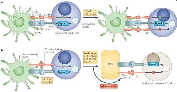
Used with permission from Macmillan Publishers Ltd: Nature Reviews Cancer, 12(4), 252-264, copyright 2012.
Future Therapeutic Targets in the Immunoglobulin Receptor Family
- There are various other activating receptors and inhibitory receptors involved in T cell activation.
- Many of these, such as TIM-3 and LAG-3 are being investigated in pre-clinical and clinical studies.
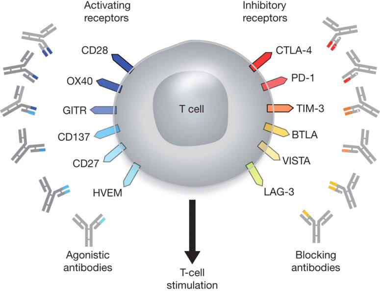
Used with permission from Macmillan Publishers Ltd: Nature, 480, 480-489, copyright 2011.
