

Immune System Overview
Components of the immune system
- The primary lymphoid organsare where lymphocytes mature.
- B cells mature in the bone marrow and T cells mature in the thymus.
- Lymphocytes that have not encountered antigen are known as naïve lymphocytes. They circulate continuously through the blood and lymphatic vessels and into the peripheral tissues.
- The secondary lymphoid organs are sites where naïve lymphocytes are activated, and include lymph nodes, spleen and mucosal-associated lymphoid tissue, such as Peyer's patches in the small intestine.
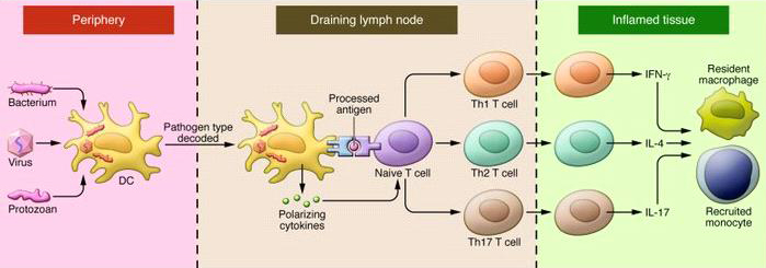
- During an infection, antigen-presenting cells (dendritic cells or macrophages) ingest pathogens or components of pathogens at the site of infection in the peripheral tissues. These peptides are referred to as antigens.
- Antigen-presenting cells travel via lymphatic vessels from the site of infection to the draining lymph nodes.
- In the lymph nodes, dendritic cells or macrophages present antigen to naive T lymphocytes, hence their name "antigen-presenting cells." In addition to presenting antigen, they provide costimulatory signals and cytokine signals.
Signals required for T cell activation
- Naïve lymphocytes receive 3 signals when they interact with antigen-presenting cells:
- An antigen signal through the T cell receptor or B cell receptor
- A co-stimulatory signal
- Naïve T cells are activated when B7.1 or B7.2 on APCs binds to CD28 on T cells.
- Naive B cells receive a co-stimulatory signal when CD40 on the B cell binds to CD40 ligand expressed on activated T cells.
- A cytokine signal

Used with permission from Macmillan Publishers Ltd: Nature Reviews Immunology, 14, 719-730, copyright 2014.
- Lymphocytes that receive all three signals from the antigen-presenting cell proliferate and differentiate into effector cells.
- B cells differentiate into plasma cells that secrete antibody, a soluble form of the B cell receptor. Antibodies bind to extracellular pathogens and toxins.
- T cells differentiate into various effector T cell subsets that either control intracellular pathogens or activate B cells to target extracellular pathogens.
- CD8 T cells become cytotoxic T cells that lyse virally-infected or tumor cells.
- CD4 T cells differentiate into various subsets of helper T cells which activate other immune cells (i.e., Th1 cells activate macrophages and Th2 cells activate B cells).
- Cells that receive signal 1 (antigen signaling through the TCR or BCR) without a co-stimulatory signal are rendered anergic, or ineffective in responding to that particular antigen.
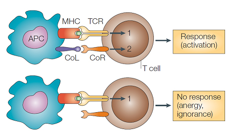
Used with permission from Macmillan Publishers Ltd: Nature Reviews Immunology, 4, 336-347, copyright 2004.
- After being activated in the secondary lymphoid organs, antigen-specific T cells migrate back to the peripheral tissue where they secrete cytokines and carry out effector functions to eliminate the pathogen that elicited the immune response.
Used with permission from the Journal of Clinical Investigation. Innate immunity in the central nervous system. Richard M. Ransohoff and Melissa A. Brown, J Clin Invest. 2012; 122(4): 1164-1171. doi:10.1172/JCI58644.
Inhibitory signals between APCs and T cells
- When lymphocytes receive the antigen and costimulatory signals from an antigen-presenting cell in the lymph node, they also upregulate inhibitory receptors.
- These inhibitory signals include CTLA-4 and PD-1, as well as many other members of the immunoglobulin family (including BTLA (B and T lymphocyte attenuator), LAG-3 (lymphocyte activation gene 3), TIM-2 (T cell immunoglobin mucin 2), A2AR (adenosine A2A receptor)).
- The purpose of these inhibitory signals is to dampen the intensity of signaling from the T cell receptor in order to prevent overactivation of the immune system that could lead to excessive tissue damage.

Used with permission from Macmillan Publishers Ltd: Nature Reviews Cancer, 12, 252-264, copyright 2012.
